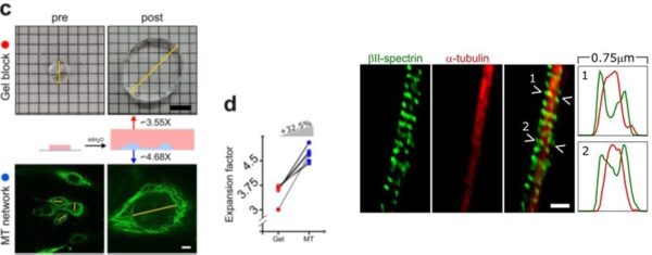
Expansion microscopy is a useful alternative to super-resolution imaging to visualize sub-diffraction cellular structures. However, it only works quantitatively if the expansion is calibrated correctly, which is not trivial. In our recent paper in Scientific Reports, led by our collaborator Dr. Nico Unsain, we present a protocol to perform expansion fluorescence microscopy quantitatively.
The expansion and calibration protocol was validated by comparing images of the membrane-associated periodic skeleton (MPS) of neurons obtained through expansion microscopy, to analogous images obtained with STED.
Scientific Reports 10 (2020) 2917
“Quantitative expansion microscopy for the characterization of the spectrin periodic skeleton of axons using fluorescence microscopy”
Gaby F. Martínez, Nahir G. Gazal, Gonzalo Quassollo, Alan M. Szalai, Esther Del Cid-Pellitero, Thomas M. Durcan, Edward A. Fon, Mariano Bisbal, Fernando D. Stefani, Nicolas Unsain


